

64,000 mirrors for sharper microscope images
Biological tissue scatters light and causes microscope images to appear blurry. Researchers at Fraunhofer IPMS have developed a chip with thousands of tiny mirrors designed to compensate for such distortions.
Read Aloud
0:00
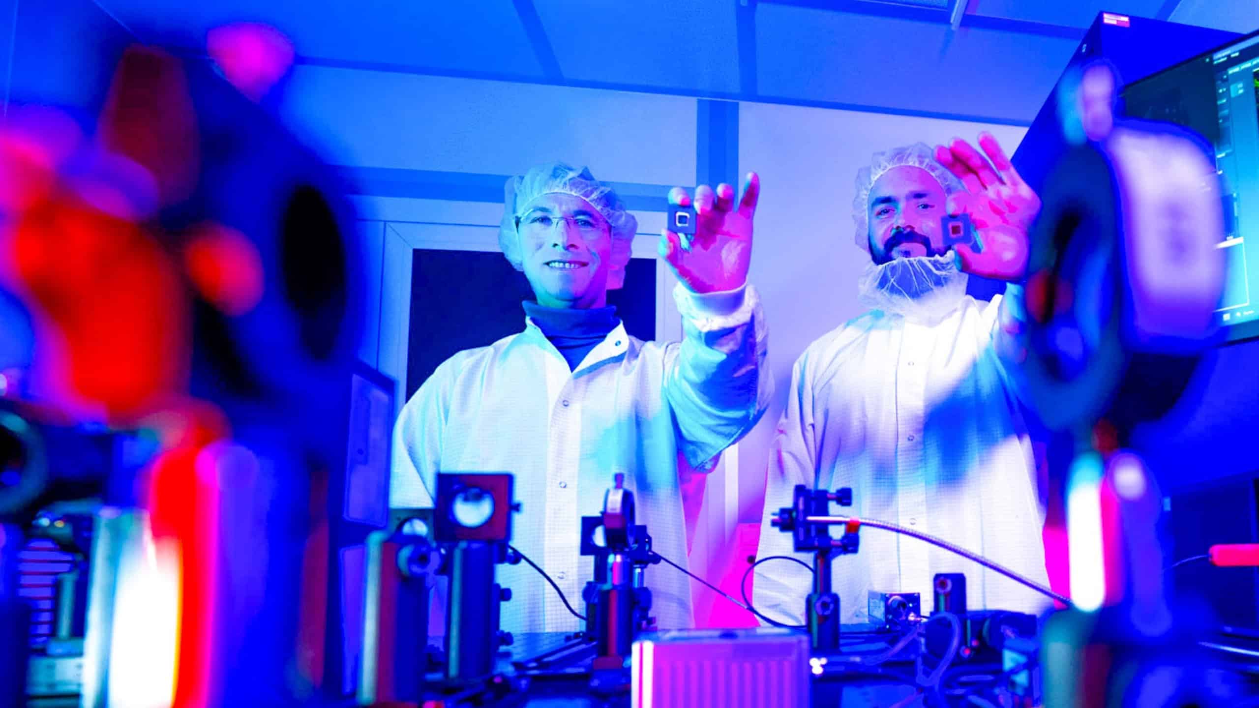
Dr. Maxim Darvin and Felix Bennewitz are conducting research on novel microspheres. The technology is designed to compensate for light distortions, thereby enabling sharper microscope images. © Sascha Thor, BTU
The deeper researchers look into tissue with a microscope, the more difficult it becomes to clearly distinguish structures. This is because the tissue scatters the light and distorts the image. Researchers at the Cottbus branch of the Fraunhofer Institute for Photonic Microsystems (IPMS) have developed a new mirror chip designed to compensate for these distortions. It could enable sharper images from deeper tissue layers in the future.
64,000 mirrors control the light
As light passes through tissue, it is scattered. The rays no longer reach the microscope as intended, and the image loses sharpness. This is particularly problematic with living tissue and when examining samples at greater depths. To compensate for such image errors, a technique called adaptive optics already exists. This involves specifically modifying the light so that a sharper image is ultimately produced. Until now, this has often required additional sensors or complicated optical systems.
“Previously, microscopes had to first measure the image aberrations and then correct them. Our new mirror array performs both tasks simultaneously – this saves time and simplifies the entire setup,” explains Dr. Maxim Darvin, project manager at Fraunhofer IPMS.
At the heart of the new technology is a chip with approximately 64,000 movable mirrors. These are so small that four of them can fit side by side within the thickness of a human hair. Each mirror can be lowered in 256 steps. By precisely adjusting the mirrors, the chip compensates for distortions in the light. The mirrors can be readjusted up to 1,000 times per second.
Correction to become faster
The researchers have already tested the technology on various biological samples. “We have already demonstrated the performance of our development on various biological samples, including corneal cells and cellulose. In all cases, our adaptive optics produced visibly sharper and higher-contrast images,” says Darvin. The team also succeeded in focusing laser light through highly scattering material. This allows structures in deeper tissue layers to be examined with higher image quality.
Although the mirrors adjust quickly, the system currently still takes a few minutes to fully determine and correct image distortions. The researchers are working to speed up this process. In the future, this should happen nearly in real time. If successful, the technology could make high-resolution microscopy faster and easier. In the long term, it could also be used in medical diagnostics.
The work is part of the OASYS research project, which runs through August 2028. The Federal Ministry of Research, Technology, and Space is funding the project with approximately 12.5 million euros. In addition to Fraunhofer IPMS, the project involves the BTU Cottbus-Senftenberg, the Ferdinand Braun Institute (FBH), and the IHP – Leibniz Institute for Innovative Microelectronics.