
The brain prepares for sight long before birth
As early as the 21st week of pregnancy, the brain is already preparing for future vision. An important structure of the visual system begins to form long before a baby opens its eyes for the first time.
Read Aloud
0:00
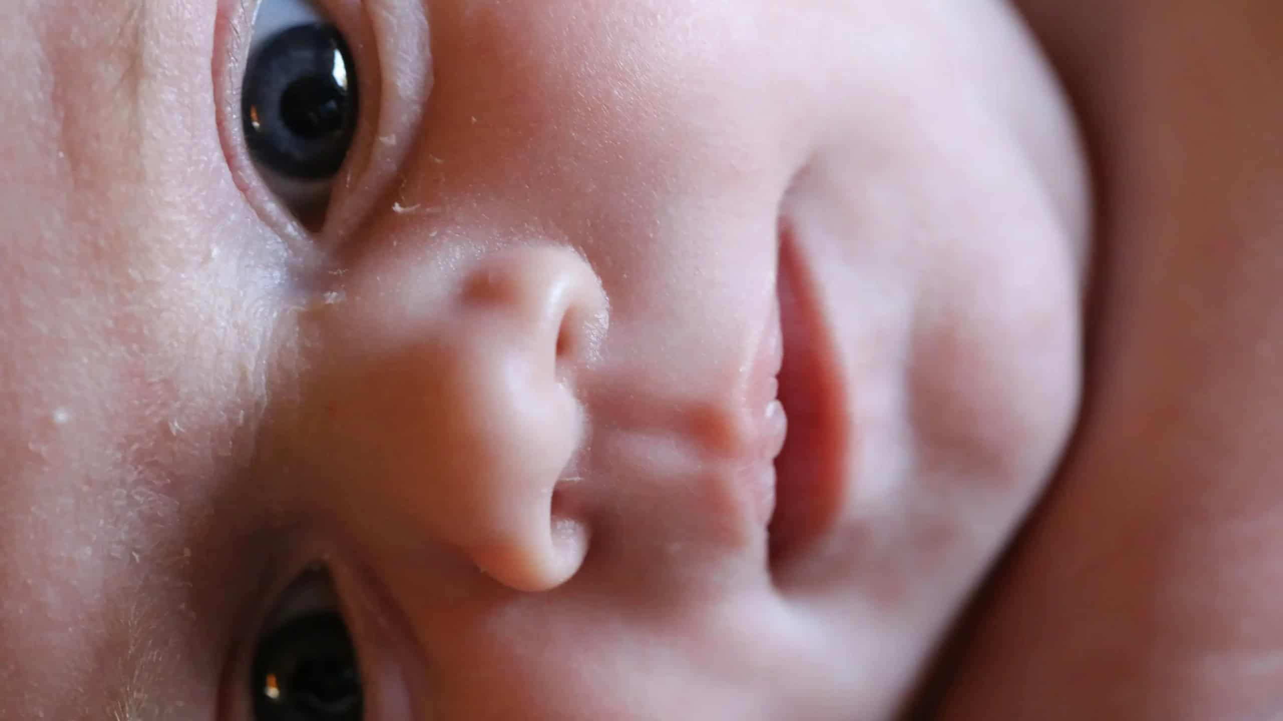
Even before birth, the brain begins preparing for vision. A research project involving scientists from Leipzig has found that the key foundations for this are already laid as early as the 21st week of pregnancy. © pixabay
A baby’s first glimpse of the world is a special moment. For the baby’s brain, however, it is less of a novelty than one might think. This is because, months before birth, a kind of “map” for future vision begins to form there – even though the child has never seen anything before. This is shown by a study in which the Max Planck Institute for Human Cognitive and Brain Sciences (MPI CBS) in Leipzig participated.
Our brain maps what we see like a map. Points that lie next to each other on the retina are also processed in the brain by neighboring nerve cells. For a long time, it was unclear whether this order arises only after birth through visual impressions or whether it already exists beforehand.
A team led by Prof. Michael Skeide investigated this question. Skeide was formerly a group leader at the MPI CBS. Today, he conducts research at the University of Kiel and at the University Medical Center Schleswig-Holstein. In Leipzig, the team developed a method that allowed them to analyze MRI data from a total of 871 fetuses, newborns, children, adolescents, and adults.
Order before the first light
The analysis shows that two key features of this “map” are already established in the brain as early as the 21st week of pregnancy. Yet fetuses do not open their eyes until around the 28th week. Furthermore, it is dark inside the uterus.
So how does this organization develop without any visual input at all?
The researchers suspect that the retina spontaneously emits regular signals even before birth. These signals could help structure the visual areas in the brain. “The results show that the basic architecture of our visual system is already established long before the first light enters the eye,” says Skeide.
To study the visual cortex in adults, researchers typically show them moving patterns while they are in an MRI scanner. This allows them to identify which areas of the brain respond to specific visual stimuli. This method does not work for unborn babies. The team therefore used MRI scans acquired without such stimuli.
These scans measure small fluctuations in blood flow in the brain, such as during sleep. From this data, the researchers were able to draw conclusions about the organization of the visual areas. The images come from the publicly accessible “Human Connectome Project.”
A look inside individual brains is still missing
So far, the results are based on the combined analysis of many brain images. Only through this approach can the weak signals in unborn infants be statistically detected. This does not yet work for a single child. Therefore, the method cannot yet be used to detect potential visual impairments at an early stage. “The next scientific step is to develop methods that allow us to reconstruct these retinal maps in the brain on an individual level,” says Skeide.
It also remains unclear how babies learn to recognize shapes and objects. “Newborns, for example, can distinguish faces shortly after birth,” the researcher emphasizes. “We find it very interesting how this is possible so early on.” Future studies will aim to clarify whether the groundwork for this is also laid before birth.
Original publication:
So-Hyeon Yoo et al., Population connective field modeling reveals proto-retinotopic visual cortex organization in the prenatal human brain. Sci. Adv. 12, eaee9300 (2026).