
Microrobot could detect ovarian cancer precursors
A robot smaller than a millimeter is designed to search for precancerous lesions in the fallopian tube and treat them directly on site. Chemnitz University of Technology is coordinating the multi-million project.
Read Aloud
0:00
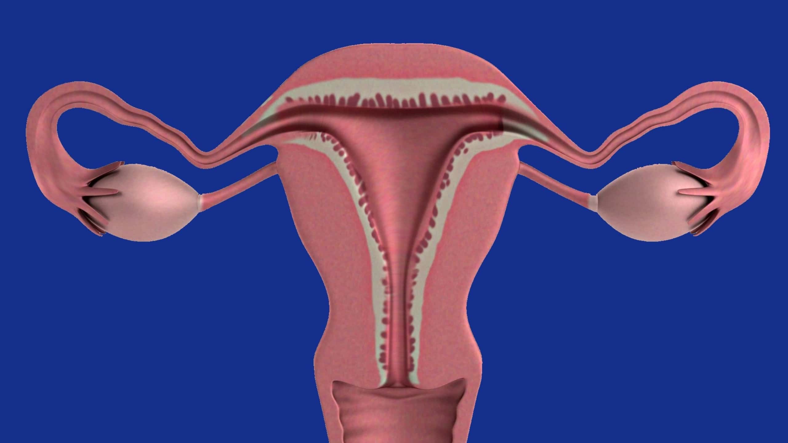
Many tumors associated with an aggressive form of ovarian cancer originate in the fallopian tube. A research team at Chemnitz University of Technology aims to develop a tiny robot that can travel through the uterus into the fallopian tube to detect precancerous lesions there. © therapractice/pixabay
The result is tiny. The robot currently being developed by a team led by Chemnitz-based researcher Oliver G. Schmidt is expected to be smaller than one millimeter. It will search the female body for early signs of an aggressive form of ovarian cancer. If everything works as planned, the microbot will also treat the developing cancer directly at the site. The European Research Council has now awarded 10.6 million euros for this project.
“Our concept involves inserting the microrobots into the fallopian tube via the uterus using a very thin catheter,” explains Oliver G. Schmidt, holder of the Chair of Material Systems for Nanoelectronics at Chemnitz University of Technology. “In principle, a major surgical procedure would not be necessary for this.”
Once the robot is in the fallopian tube, it will be guided from the outside using magnetic fields. Ultrasound provides images from inside the body and simultaneously serves as a power source for the mini-bot. With the help of artificial intelligence, the images can be used to precisely track where the robot is and how it is moving. If it detects the characteristics of a precancerous lesion, it releases an active ingredient at the affected site.

Schmidt and his team, together with researchers from the universities of Manchester and Oxford, aim to investigate whether this plan can be implemented. They have named their project ECLIPSE. It will receive funding for the next six years. For Chemnitz University of Technology, which is coordinating the project, this is the first so-called ERC Synergy Grant from the European Research Council.
Why the search begins in the fallopian tube
High-grade serous ovarian carcinoma is an aggressive form of ovarian cancer. It is often detected very late – usually only after it has already spread. A nationwide analysis of nearly 39,000 cases with known tumor stage illustrates this. In about three out of four patients, the disease was already advanced at the time of diagnosis. If the cancer was detected at the earliest stage, the five-year survival rate was about 86 percent. At the most advanced stage, the survival rate was around 30 percent.
Many of these tumors originate in the fallopian tubes. Over the course of years, tiny foci of cancer develop there. They are difficult to reach in the narrow fallopian tubes using standard examination methods. “Our partners in Manchester have been studying the development of ovarian cancer for years and have found evidence that very early precancerous lesions in the fallopian tube can be detected based on specific molecular signals,” reports Schmidt. However, a single signal is often not enough to reliably detect these early signs.
Together with the team in Oxford, the researchers therefore plan to combine several markers. “The robot should only trigger a therapeutic response when several precisely defined conditions are met simultaneously.” This would prevent the robot from releasing an active ingredient in response to other changes in the tissue. But the road ahead is challenging. The project aims to determine whether the microrobot can reliably detect precancerous lesions and which active ingredients are suitable for treatment.
Morphobots change their shape
Schmidt’s research group is contributing its expertise in micro-robotics to this effort. They have already developed models that are smaller than the diameter of a human hair. “The key is determining which functions can be integrated into the smallest possible space,” explains the professor. This year, the Chemnitz team unveiled their so-called “Smartlets.” These tiny robots carry electronic chips. They can harvest energy from their environment, process information, communicate with one another, and assemble into larger units.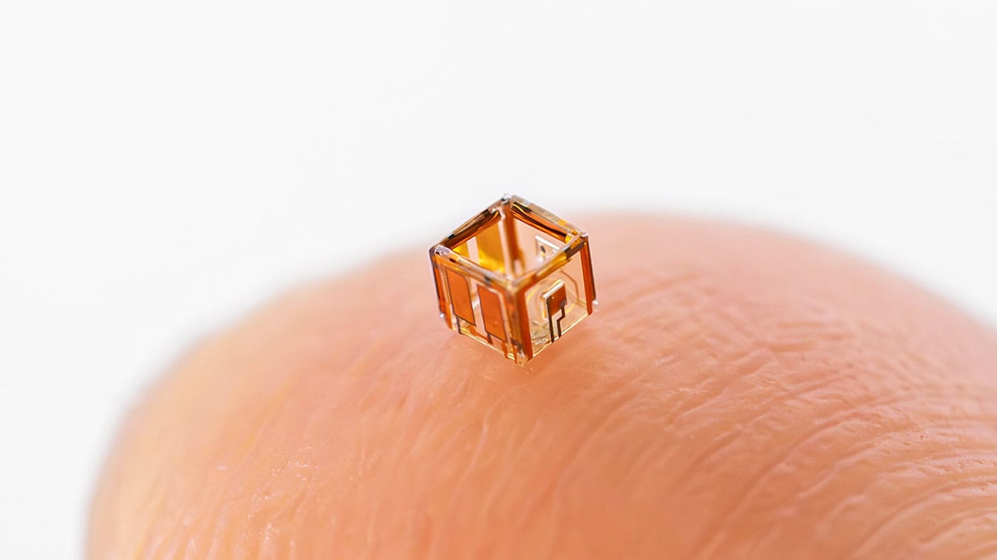
The robots designed for the fallopian tube now present a new challenge. They are also intended to detect biological signals and release the active ingredient. “We call this new class of microrobots ‘Morphobots’ because they are designed to be able to change their shape in a targeted manner.” Another question for the coming years is how to remove these tiny helpers after they’ve completed their mission. “We must ensure that the robots do not damage the sensitive tissue and do not pose a risk even if their movement or control does not function as intended.”
Navigating through narrow structures in the body, detecting cancer signals, and ultimately releasing the active ingredient – the list of tasks for the next six years is long. By the end of the project, the partners aim to demonstrate that a single microrobot can accomplish all of this. This will be tested using human tissue and in animal models.
“If, at the end of the six years, we can demonstrate that such an integrated approach works in principle and also meets the necessary safety requirements, that would be a significant scientific breakthrough,” says Schmidt, outlining the goal. Clinical trials with patients could then be prepared.
Later, microrobots could also be deployed in other parts of the body to detect diseases – in places that have been difficult to examine using conventional methods.